Original Story
Scientists Found Preserved Blood Vessels Inside a T. Rex Fossil. The Dinosaur Had Been Hurt Before It Died.
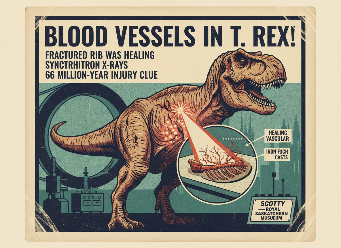
Inside a fossilized rib from the largest Tyrannosaurus rex ever discovered, researchers have found something they did not expect: an intricate network of iron-rich structures that preserve the shape and pattern of the blood vessels that were present when the bone was healing, 66 million years ago. The discovery was published in Scientific Reports and announced April 26, 2026. The T. rex in question is nicknamed Scotty, housed at the Royal Saskatchewan Museum in Canada. Scotty appears to have had a rough life. Its bones show multiple signs of injury, including at least one fractured rib that was in the process of healing but never finished. The new study used synchrotron radiation — an extremely powerful type of X-ray generated by a particle accelerator — to peer inside the dense fossil bone without damaging it, and found what appears to be a cast of the original blood vessel network, preserved in mineralized iron, left behind by the dinosaur’s own healing process.
Finding soft tissue evidence inside a dinosaur fossil is rare enough to be significant under any circumstances. Dinosaurs lived between 245 and 66 million years ago. Over that kind of time, the organic material in bone — the cells, proteins, blood vessels, and other soft structures — normally decays completely, leaving only the mineralized portions behind. This is why paleontologists typically have access to nothing but bones and teeth: those are the structures hard enough to resist decomposition across geological time.
In rare cases, exceptional preservation conditions can protect soft tissue evidence. Amber preserves insects in remarkable detail. Permafrost can preserve skin, hair, and even gut contents in Ice Age mammals. In fossilized bones, scientists have occasionally found traces of original organic material, including fragments of protein and, in a handful of disputed cases, traces that some researchers argue may be original soft tissue. What the Scotty rib provides is something more specific: not disputed fragments, but a clear three-dimensional network of mineralized structures whose shape, branching pattern, and density match what you would expect from blood vessels in healing bone.
Why a Fractured Rib Is the Key
The preservation happened because the rib was injured. When an animal breaks a bone, the body’s immediate response is to dramatically increase blood flow to the damaged area. Specialized blood vessels proliferate rapidly in the injured tissue, delivering the raw materials needed for bone repair — calcium, phosphate, and the cellular machinery to assemble them. In living animals, this process produces a distinctive tangle of small vessels woven through the healing tissue, visible in cross-sections of the bone.
In Scotty’s rib, that healing process was underway but incomplete when the animal died. The bone fracture shows evidence of partial repair, but the rib was never fully restored to its original integrity. This means Scotty died sometime after the injury — possibly weeks or months later — while the healing network of blood vessels was still present in the bone tissue.
Over the following 66 million years, the iron from the hemoglobin in those blood vessels concentrated in the tissues as the organic material decayed. The iron left behind mineralized casts of the original vessel shapes — hollow tubes filled with iron oxide, preserving the path and branch structure of the vascular network in a form that synchrotron X-rays could map in three dimensions.
Lead researcher Iris Mentz, a physics PhD student at the University of Regina who joined the research team as an undergraduate, explained the significance: “By analyzing blood vessels produced by an incompletely healed fracture, we can hopefully learn how T. rex healed, helping speculation on how Scotty was able to survive after sustaining injuries.”
What This Means Beyond Scotty
The implications go in two directions. The first is biological: for the first time, researchers have a structural model of the healing blood vessels of a T. rex, which they can compare to equivalent structures in modern birds and crocodilians — both of which are evolutionary relatives of dinosaurs — to test ideas about how dinosaur physiology resembled or differed from its modern descendants.
The second implication is methodological. Synchrotron scanning does not damage the specimen. It can be applied to fossils that would be destroyed by conventional dissection, and it can penetrate the dense mineral matrix of fossilized bone that blocks standard CT scanners. The Scotty findings suggest that other injured dinosaur bones — specimens that were previously passed over as damaged or pathological rather than scientifically useful — may contain preserved vascular networks that have simply never been looked for with the right technology.
As Mentz noted, scientists may need to rethink which fossils are most valuable. A broken bone may have more to say than a whole one.
Sources: Scientific Reports — Mentz et al., Blood Vessel Preservation in a T. rex Rib: Synchrotron Analysis of Healing Vascular Structures in a Fractured Fossil Bone (April 26, 2026) — ScienceDaily — Blood Vessels Found in T. Rex Bones Are Rewriting Dinosaur Science (April 26, 2026) — Colombia One — T. Rex Blood Vessels Discovery Reveals How Dinosaurs Healed Injuries (April 27, 2026) — Greek Reporter — Scientists Discover Preserved Blood Vessels in T. Rex Dinosaur Fossil (April 28, 2026) — Animals Around the Globe — Blood Vessels Found in T. Rex Bone Unlock Secrets of Dinosaur Biology (April 26, 2026) — SciTechDaily — Blood Vessels Found in T. Rex Bones Rewrite What We Know About Dinosaurs
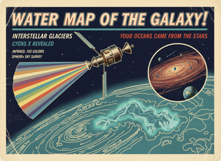
NASA Has Mapped the Water in the Galaxy. The Ocean Beneath Your Feet Came From Out There.
A study published in The Astrophysical Journal on April 15, 2026, based on data from NASA's SPHEREx mission, has produced the first…
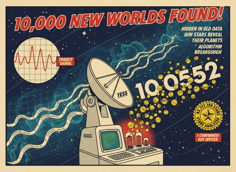
A Machine Learning Algorithm Just Found 10,000 Alien Planets Nobody Had Seen Before. They Were Hidden in Data We Already Had.
The current official count of confirmed exoplanets — planets orbiting stars other than our sun — sits at roughly 6,300. It took…
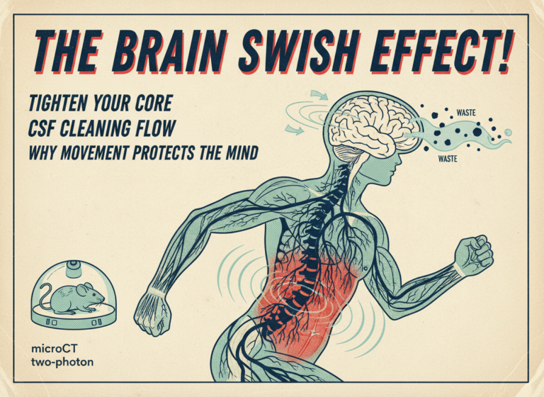
Every Time You Tighten Your Core, Your Brain Gently Swishes Around. That Might Be Keeping You Sane.
When you tighten your abdominal muscles — whether you are walking, standing up from a chair, or doing anything that engages your…
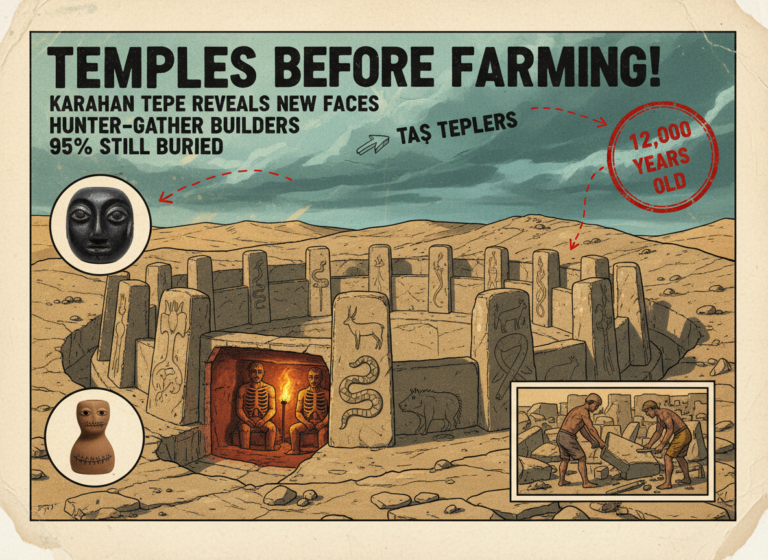
They Built a Temple Complex Before They Had Farming, Writing, or Pottery. New Finds at Karahan Tepe Just Made the Mystery Bigger.
Fresh discoveries at a 12,000-year-old site in Turkey called Karahan Tepe, the possible earliest village in human history, have uncovered more carved…